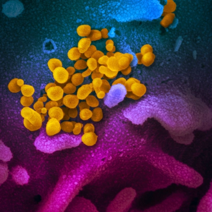
Last reviewed: January 5, 2022
The following is a curated review of key information and literature about this topic. It is not comprehensive of all data related to this subject.
Overview
Clinicians typically diagnose respiratory infection by viruses such as SARS-CoV-2 through direct detection of viral nucleic acid or protein in respiratory tract specimens. The two most frequently used tools to do this are nucleic acid amplification tests such as the polymerase chain reaction and rapid antigen-based tests. When the COVID-19 pandemic began, reverse-transcriptase PCR tests were the first to be developed and widely deployed.
NAATs such as RT-PCR for SARS-CoV-2 are designed to detect viral RNA. A positive result is highly specific for the presence of viral nucleic acid; however, it does not differentiate between viable and nonviable virus. Thus, a positive test does not necessarily indicate that a person is infectious and requires isolation.
A variety of gene targets are available across different assays, including the envelope gene, the nucleocapsid gene, the ORF1ab gene and the spike gene. Most assays are designed to detect two or more gene targets. FDA maintains an up-to-date list of assays with emergency use authorization.
An NAAT’s clinical sensitivity is affected by the assay’s limit of detection, the time since infection began and sample type. Because patients with symptoms tend to present earlier in their infection course, there is increased test sensitivity in symptomatic people relative to asymptomatic people. Traditionally the nasopharynx has been the preferred body site for sampling, but given patient discomfort and the need for serial testing, many hospitals and public health programs now use anterior nares or saliva as the primary sampling type.
Here, we focus on literature related to factors affecting molecular SARS-CoV-2 diagnostics’ clinical sensitivity, which include the dynamics of viral replication in natural infection, the impact of alternative sample sites and the implications of persistent positivity.
Guidelines
IDSA guidelines recommend NAAT (including RT-PCR) for:
- All symptomatic individuals suspected of having COVID-19;
- Asymptomatic individuals with known or suspected contact with a COVID-19 case;
- Asymptomatic individuals when the results will impact isolation/quarantine/personal protective equipment usage decisions, dictate eligibility for surgery or inform administration of immunosuppressive therapy.
Additionally, IDSA recommends:
- NAAT testing in symptomatic people, even when clinical suspicion is low;
- NAAT testing in asymptomatic individuals with known or suspected exposure to COVID-19, those requiring hospitalization when community prevalence is high or prior to solid organ or stem cell transplantation and time-sensitive surgeries;
- Repeating viral RNA testing when the initial test is negative in symptomatic individuals with intermediate or high clinical suspicion;
- Specimen collection from the nasopharynx, mid-turbinate, anterior nares or combined nasal/oropharyngeal over oropharynx alone.
Key Literature
In summary: Overall, NAAT is the mainstay in diagnosing COVID-19. Consider timing of NAAT testing in relation to onset of infection, assay limit of detection, sample collection site location and the detection of nonviable virus. Innovations in NAAT include improved algorithms for handling false negative or false positive results, pooling, faster turnaround times and reductions in the use of materials.
Duration of infectiousness and correlation with RT-PCR cycle threshold values in cases of COVID-19 (Singanayagam, August 2020).
Overall, in this retrospective study of samples taken for national surveillance purposes, SARS-CoV-2 viral load in the upper respiratory tract peaked around the time of symptom onset. The ability to culture SARS-CoV-2 in patients with mild-moderate disease was highest in week one, and declined significantly by day 10 after symptom onset; by day 12 virus was unable to be cultured. RT-PCR cycle threshold values correlated strongly with cultivable virus.
Study population:
- 754 upper respiratory tract (URT) samples from 425 people with symptomatic COVID-19 tested at Public Health England’s national respiratory virus reference laboratory between January and April 2020.
- Samples included nose, throat, combined nose-and-throat and nasopharyngeal swabs, or nasopharyngeal aspirates.
- Virus culture was attempted on 253 cases; 233 cases (92%) were classified as non-severe (asymptomatic or mild-to-moderate) and 20 (8%) had severe illness.
Primary endpoint:
- To determine the duration of infectiousness and determine if RT-PCR detection relates to cultivable virus.
Key findings:
- In the first week after symptom onset (days −2 to 7), mean cycle threshold (Ct) was 28.18 (95% CI 27.76–28.61); in the second week Ct was 30.65 (95% CI: 29.82–31.52). After 14 days, geometric mean (GM) Ct was 31.60 (95% CI: 31.60–34.49).
- There was no difference in Ct values between those with mild-to-moderate and severe disease (p = 0.79).
- There was no difference in culture positivity between symptomatic and asymptomatic cases (OR = 0.66; 95% CI: 0.34–1.31).
- The estimated probability of recovery of virus from samples with Ct > 35 was 8.3% (95% CI: 2.8%–18.4%).
- There were 246 samples from 176 symptomatic cases where the date of symptom onset was known: of these, 103 (42%) from 81 cases were culture-positive.
- The culture positivity rate was significantly higher during week 1 than week 2 (74% vs 20%; p = 0.002).
- Ten days after symptom onset, the probability of culturing virus declined to 6.0% (95% CI: 0.9–31.2%)
- After day 12, virus was not cultured.
Limitations:
- Patients’ recollection was used to determine symptom onset; recall bias is possible.
- Duration and cessation of symptoms was not well recorded.
- Most cases in which culture was attempted were mild-moderate; this may limit the generalizability of the findings to severe disease.
- For asymptomatic cases, the time of infection acquisition was not known.
- Subjects were not sampled systematically, which could have caused bias in the timing of sampling related to the clinical scenario.
Salivary Detection of COVID-19 (Caulley, August 2020).
Overall, in this prospective study, saliva testing with a SARS-CoV-2 RT-PCR was less sensitive than nasopharyngeal or oropharyngeal swab testing.
Study population:
- Asymptomatic, high-risk individuals and people with mild symptoms suggestive of COVID-19 at a testing center in Canada (n=1939 paired samples of either nasopharyngeal or oropharyngeal swabs, and saliva).
Primary endpoint:
- To determine the detection rate of SARS-CoV-2 using a saliva specimen compared with standard swab testing.
Key findings:
- SARS-CoV-2 was detected in 70 samples - 80.0% with swabs and 68.6% with saliva.
- 34 patients (48.6%) tested positive for SARS-CoV-2 on both swab and saliva samples.
- 22 patients (31.4%) tested positive with swab alone and 14 (20%) tested positive with saliva alone.
Limitations:
- Swab and saliva samples were split between 2 laboratories, which may have led to reporting bias.
- The sample size of patients with COVID-19 was small (n=34 patients).
Prospective Saliva or Nasopharyngeal Swab Specimens for Detection of SARS-CoV-2 (Wyllie, August 2020).
Overall, in this prospective study, saliva specimen was associated with similar sensitivity to nasopharyngeal swab in detecting SARS-CoV-2 RNA.
Study population:
- 70 hospitalized patients with COVID-19, confirmed by a positive nasopharyngeal swab specimen.
- After diagnosis, patients also provided saliva samples.
Primary endpoint:
- To assess how saliva compares with nasopharyngeal swab specimens with respect to sensitivity in detection of SARS-CoV-2 during the course of infection.
Key findings:
- More SARS-CoV-2 RNA copies in the saliva specimens were found (mean log copies per milliliter, 5.58; 95% CI, 5.09-6.07) than in the nasopharyngeal swabs (mean log copies per milliliter, 4.93; 95% CI, 4.53-5.33).
- At 1 to 5 days after diagnosis, 81% (95% CI, 71-96) of the saliva samples were positive, compared with 71% (95% CI, 67-94) of the nasopharyngeal swab specimens.
- Less variation in levels of SARS-CoV-2 RNA was observed in the saliva specimens (standard deviation, 0.98 virus RNA copies per milliliter; 95% credible interval, 0.08 to 1.98) than in the nasopharyngeal swab specimens (standard deviation, 2.01 virus RNA copies per milliliter; 95% credible interval, 1.29 to 2.70).
Limitations:
- Small sample size of individuals that had confirmatory COVID-19.
- Single-center study.
- It appears that 100% of patients with positive nasopharyngeal swabs also had positive saliva samples, but this is not clearly stated in the manuscript; if accurate, would be a better performance than other papers studying the sensitivity of saliva SARS-CoV-2 testing.
Prospective Evaluation of Saliva as a Noninvasive Specimen for Detection of SARS-CoV-2 (Williams, July 2020).
Overall, in this prospective study assessing the feasibility of saliva specimens, a majority of patients with positive nasopharyngeal swabs for SARS-CoV-2 also had positive saliva samples, but the sensitivity of the latter test was lower. Higher viral loads were noted with nasopharyngeal swabs.
Study population:
- 622 patients at a COVID-19 screening clinic in Australia with positive nasopharyngeal RT-PCRs for SARS-CoV-2.
- 522 (83.9%) also provided saliva specimens.
Primary endpoint:
- To assess the feasibility of saliva specimens in comparison to nasopharyngeal swabs in detecting SARS-CoV-2.
Key findings:
- 39/622 (6.3%; 95% CI, 4.6-8.5%) patients had PCR-positive nasopharyngeal swabs; of these, 33/39 patients (84.6%; 95% CI, 70.0-93.1%) had SARS-CoV-2 detected in saliva.
- The median cycle threshold (CT) was significantly lower in NPS compared to saliva, suggesting higher viral loads in the nasopharynx.
- In both nasopharyngeal and saliva samples, there was a correlation between CT value and days from symptom onset.
Limitations:
- Small sample size of patients with positive nasopharyngeal swabs (n=39).
- No information was provided regarded patients’ baseline characteristics and symptoms; severity of COVID-19 was not reported.
Variation in False-Negative Rate of Reverse Transcriptase Polymerase Chain Reaction–Based SARS-CoV-2 Tests by Time Since Exposure (Kucirka, May 2020).
Overall, in this pooled analysis, false negative RT-PCRs were least common 3 days after symptom onset. The rate of false-negative RT-PCRs was highest the day of infection, were lowest 8 days after infection, and then began to rise again.
Study population:
- Pooled analysis of a mix of inpatients and outpatients with SARS-CoV-2 infection in 7 studies of RT-PCR performance in the upper respiratory tract by time since symptom onset or exposure (n = 1330 respiratory samples).
Primary endpoint:
- Estimation of false-negative rates by day since infection.
Key findings:
- Over the 4 days between infection (day 1) to the typical time of symptom onset (day 5), the probability of a false-negative result in an infected person decreased from 100% on day 1 (95% CI, 100%-100%) to 67% (CI, 27% - 94%) on day 4.
- On the day of symptom onset (day 5), the median false-negative rate was 38% (CI, 18% to 65%).
- On day 8 the median false-negative rate decreased to 20% (CI, 12%- 30%), and then began to increase again (21% [CI 13%-31%] on day 9).
- On day 21 the median false negative rate was 66% (CI, 54% -77%).
Limitations:
- Analysis based on heterogenous studies.
- Authors assumed date of infection was 5 days before symptoms began; possible this estimate was inaccurate for some patients.
- Sample collection techniques varied across studies.
- It was not possible to separate false-negative rates by sample type (oropharyngeal vs. nasopharyngeal); this may have different false negative proportions.
Predicting Infectious Severe Acute Respiratory Syndrome Coronavirus 2 From Diagnostic Samples (Bullard, May 2020).
Overall, in this retrospective cross-sectional study, the ability to culture SARS-CoV-2 at a Ct > 24 or duration of symptoms > 8 days may be low. The authors assume culture is an adequate surrogate for infectivity; while this is a reasonable assumption, it has not been proven.
Study population:
- Retrospective cross-sectional study of 90 patients with COVID-19 in Canada.
- Nasopharyngeal and endotracheal samples obtained from persons tested between 0-21 days after symptom onset and incubated on Vero cells.
Primary endpoint:
- The relationship between SARS-CoV-2 RT-PCR cycle threshold (Ct) values from respiratory samples, symptom onset to test (STT), and infectivity in cell culture.
Key findings:
- Twenty-six samples (28.9%) demonstrated viral growth.
- No growth was observed in samples with a Ct > 24 or STT of > 8 days.
- Ct was found to be statistically associated with positive culture (OR 0.64 [95% CI: 0.49 – 0.84]); for each 1 unit increase in Ct value the odds of infectivity decreased by 32%.
- STT was associated with positive culture (OR 0.63 [95% CI: 0.42 – 0.94]); For every 1-day increase in STT, the odds ratio of being culture positive was decreased by 37%
- The peak probability of a positive culture was on day 3 STT and decreased thereafter.
Limitations:
- Relatively small sample size; not clear how many samples were available by day.
- No patients were asymptomatic; these results may not be generalizable to this population.
- Patient recollection was used to determine symptom onset; recall bias is possible.
Saliva sample as a non-invasive specimen for the diagnosis of coronavirus disease 2019: a cross-sectional study (Pasomsub, May 2020).
Overall, in this cross-sectional study, saliva sample was associated with a strong agreement of SARS-CoV-2 RT-PCR positivity compared with nasopharyngeal and throat swabs; however, the small sample size and inclusion of only symptomatic patients limits generalizability of the findings.
Study population:
- 200 individuals under investigation who attended an acute respiratory infection clinic in Thailand.
- Individuals were included if they had a travel history from an endemic area of COVID-19 within 14 days or had a history of contact with an individual who was confirmed to have or suspected of having COVID-19.
- Individuals provided nasopharyngeal swabs, throat swabs, and saliva samples
Primary endpoint:
- To determine the feasibility of saliva specimens to detect SARS-CoV-2.
Key findings:
- Among 19 patients diagnosed with COVID-19 by nasopharyngeal and throat swab RT-PCR, the median (IQR) onset of symptoms before the test was 3 (2–11) days.
- Using nasopharyngeal and throat swab RT-PCR as the reference standard, the prevalence of COVID-19 diagnosed by nasopharyngeal and throat swab RT-PCR was 9.5% (19/200) compared to 9% (18/200) with saliva.
- The sensitivity and specificity of the saliva samples were 84.2% (95% CI 60.4–96.6%), and 98.9% (95% CI 96.1–99.9%), respectively.
- Positive predictive value and negative predictive value were 88.9% (95% CI 65.3%–98.6%), and 98.4% (95% CI 95.3%–99.7%).
- An analysis of the agreement between nasopharyngeal and throat compared to saliva showed a 97.5% observed agreement (κ coefficient 0.851; p < 0.001).
Limitations:
- Only patients with respiratory symptoms were enrolled; therefore, these results cannot be applied to asymptomatic patients.
- Small sample size (n=19).
Virological assessment of hospitalized patients with COVID-19 (Wölfel, April 2020).
Overall, in this small study of patients with mild COVID-19, peak virus load via RT-PCR was observed early after symptom onset (within 4 days) and viable virus was not cultured past day 8.
Study population:
- 9 hospitalized patients with mild COVID-19 from an epidemiologic cluster in Germany.
- All patients were initially diagnosed by RT–PCR from oro- or nasopharyngeal swab specimens.
Primary endpoint:
- Viral load in patients with COVID-19.
Key findings:
- The average viral load by RT-PCR was 6.76 × 10⁵ copies per whole swab until day 5; viral load peaked on day 4, with 7.11 × 10⁸ RNA copies per throat swab.
- Samples taken after day 5 had an average viral load of 3.44 × 10⁵ copies per swab.
- SARS-CoV-2 was able to be cultured during the first week of symptoms in 16.66% of swabs and 83.33% of sputum samples.
- Although viral RNA was detected by RT-PCR up to 28 days after symptom onset, viable virus was not able to be cultured after 8 days.
- Once RT-PCR dropped below 106 copies/mL virus is no longer culturable.
- Seroconversion in 50% of patients occurred by day 7, and in all patients by day 14, but this did not correlate with a rapid decline in viral load.
Limitations:
- Small sample size.
- All patients had mild COVID-19, which may limit the generalizability of findings to patients with moderate-severe disease.
Presymptomatic SARS-CoV-2 Infections and Transmission in a Skilled Nursing Facility (Kimball, April 2020).
Overall, in this prospective point prevalence study of skilled nursing residents, patients who were presymptomatic had high viral loads by RT-PCR.
Study population:
- 76 residents in a skilled nursing facility (48 positive for COVID-19).
- Residents were categorized as symptomatic if they had had at least one new or worsened typical or atypical symptom of COVID-19 in the preceding 14 days.
- Asymptomatic residents were those who had no symptoms or only stable chronic symptoms (e.g., chronic cough without worsening).
- Presymptomatic residents were those who were asymptomatic at the time of testing but developed symptoms within 7 days after testing.
Primary endpoint:
- Transmission of SARS-CoV-2 and the adequacy of symptom-based screening to identify infections in residents.
Key findings:
- 27 of 48 residents (56%) were asymptomatic at the time of testing; of these patients, 24 (88.9%) developed symptoms.
- Samples from these 24 presymptomatic residents had a median rRT-PCR cycle threshold value of 23.1, indicating a high viral load
- Viable virus was cultured from 17 residents.
Limitations:
- This analysis was conducted in skilled nursing residents, which may limit the generalizability of the findings.
- Symptom ascertainment in population was difficult, which could have lead to misclassification of patients.
Temporal dynamics in viral shedding and transmissibility of COVID-19 (He, April 2020).
Overall, in this prospective study, viral load in patients with COVID-19 was highest at the time of symptom onset.
Study population:
- 94 hospitalized patients with COVID-19 in China.
- 66% were moderately ill; none were classified as ‘severe’ or ‘critical’ on hospital admission.
- 414 throat swabs were collected, from symptom onset up to 32 days after onset.
- Modeled COVID-19 infectiousness profiles from a separate sample of 77 infector–infectee transmission pairs.
Primary endpoint:
- Temporal patterns of viral shedding.
Key findings:
- High viral loads were detected soon after symptom onset, then gradually decreased towards the detection limit by about day 21.
- There was no difference in viral loads across sex, age groups and disease severity.
- Based upon modeling COVID-19 infectiousness profiles, 44% of secondary cases were infected during the index cases’ presymptomatic stage (95% CI 30–57%).
Limitations:
- Patient recollection was used to determine symptom onset; recall bias is possible.
- Most patients had moderate COVID-19, which may limit the generalizability of these findings to patients with severe disease.
Viral load dynamics and disease severity in patients infected with SARS-CoV-2 in Zhejiang province, China, January-March 2020: A retrospective cohort study (Zheng, April 2020).
Overall, in this retrospective cohort study the median duration of positive RT-PCR tests in respiratory samples was 21 days in patients with severe disease and 14 days in patients with mild disease. The duration of SARS-CoV-2 positivity by RT-PCR was significantly longer in stool samples than in respiratory and serum samples.
Study population:
- Retrospective study of 96 hospitalized patients with COVID-19 in China; 22 with mild disease and 74 with severe disease.
- 3497 respiratory, stool, serum, and urine samples were collected.
Primary endpoint:
- Viral load by RT-PCR at different stages of COVID-19 disease.
Key findings:
- RNA was detected in the stool of 55 patients (59%) and in the serum of 39 patients (41%).
- Median duration of virus in stool (22 days, IQR 17-31 days) was significantly longer than in respiratory (18 days, IQR 13-29 days; P=0.02) and serum samples (16 days, IQR 11-21 days; P<0.001).
- Median duration of virus in the respiratory samples of patients with severe disease (21 days, IQR 14-30 days) was significantly longer than in patients with mild disease (14 days, IQR 10-21 days; P=0.04).
- In patients with mild disease, viral loads peaked in respiratory samples in the second week from disease onset; in patients with severe disease, viral load continued to be high during the third week of disease.
Limitations:
- Single-center study.
- Data collected retrospectively, which may have allowed for confounding.
- A small number of patients in the sample (22) had only mild disease.
Prospective Evaluation of SARS-CoV-2 Viral Load in Upper Respiratory Specimens of Infected Patients (Zou, March 2020).
Overall, in this small prospective study of patients with COVID-19, high viral loads were present early after symptom onset, compared to later.
Study population:
- 18 patients within 2 family clusters with COVID-19 from China - 1 person was asymptomatic, while 13 had pneumonia on CT.
- 72 throat swabs and 72 nasal swabs were collected.
Primary endpoint:
- Viral load measurements via RT-PCR.
Key findings:
- Viral loads were higher soon after symptom onset compared to later.
- Higher viral loads were detected in the nose compared to the throat of patients.
- The viral shedding pattern resembled that of influenza rather than SARS-CoV-2.
- The viral load of the asymptomatic patient was similar to that of the symptomatic patients.
Limitations:
- Small sample size.
- Patients were part of the same family cluster; viral inoculum may have been higher in this group than in patients who are infected outside the home.
Retrospective Evaluation of Detection of SARS-CoV-2 via RT-PCR in Different Types of Clinical Specimens (Wang, March 2020).
Overall, in this retrospective evaluation of SARS-CoV-2 RT-PCR from various sites, samples from the lower respiratory tract had higher positivity rates than samples from the upper respiratory tract. Viral loads were higher in the nares.
Study population:
- 1070 specimens of SARS-CoV-2 from different sites in 205 hospitalized patients with COVID-19, 19% of whom had severe illness.
- RT-PCR pharyngeal swabs were collected from most patients 1 to 3 days after hospital admission.
- Blood, sputum, feces, urine, and nasal RT-PCR samples were collected throughout hospitalization.
- Bronchoalveolar lavage fluid and fibrobronchoscope brush biopsy were sampled from patients with severe illness or undergoing mechanical ventilation.
Primary endpoint:
- To determine the biodistribution of SARS-CoV-2 in various tissues.
Key findings:
- Bronchoalveolar lavage fluid specimens showed the highest positive rates (14/15; 93%), followed by sputum (72/104; 72%), nasal swabs (5/8; 63%), fibrobronchoscope brush biopsy (6/13; 46%), pharyngeal swabs (126/398; 32%), feces (44/153; 29%), and blood (3/307; 1%).
- Nasal swabs had a higher mean cycle threshold than other sites (24.3, [1.4 × 10⁶ copies/mL] vs 30 [(2.6 × 10⁴ copies/mL]), indicating higher viral loads in the nares.
Limitations:
- Some patients did not have detailed clinical information available, limiting the data from being correlated with symptoms or disease course.
- The sample size was small from some tissue areas.
- The timing to sampling differed between sites, and thus the results may not be directly comparable.
- Lower respiratory tract sampling occurred only in patients with severe disease; findings for these patients may not be directly comparable with those for patients with mild-moderate disease.
Additional Literature
Evaluating the accuracy of different respiratory specimens in the laboratory diagnosis and monitoring the viral shedding of 2019-nCoV infections (Yang, February 2020). 213 patients hospitalized with COVID-19 in China provided 866 clinical respiratory samples, including nasal and throat swabs, sputum, and bronchoalveolar lavage fluid. Sputum samples were most likely to test positive by RT-PCR, regardless of the clinical severity of disease.
Detection profile of SARS-CoV-2 using RT-PCR in different types of clinical specimens: A systematic review and meta-analysis (Bwire, July 2020). 8136 pooled clinical specimens analyzed to detect SARS-CoV-2; most were nasopharyngeal swabs (69.6%). Lower respiratory tract specimens had a positive rate of 71.3%; bronchoalveolar lavage fluid had a positive rate of 91.8%; sputum had a positive rate of 68.1%; and nasopharyngeal swabs had a positive rate of 45.5%.